


In the average week, we have many cases of eye infections and conditions that we often document to show progress.
First, about the camera...
Attached to my biomicroscope is a small digital image capturing camera. There is no zoom and lighting is VERY difficult. Part of the reason why is that to get these shots, you often times have to really brighten the light and you risk putting an uncomfortable patient in greater discomfort. Since we capture these shots for insurance purposes, we dont need to get a "perfect" shot.....but the photographer in me WANTS a good shot. IIRC the resolution of these images is 2.8mp....the image capture system is about 7 years old...
ONe other thing....as I reviewed these images, I noticed that you might all see a bright white reflection. These are usually artifact and a result of reflection off the cornea. So I will do my best to educate and describe what you are looking at. Any questions please ask.
OK, here's a fraction of a day in the office....not sure if it meets the definition of macro though.
But I am hoping you'll find this interesting as the eyes are the ONLY CAMERAS THAT WE ALL HAVE IN COMMON!!
The cornea is the front windshield of the eye....The majority are at the corneal surface and I will state otherwise if not...
A perfectly normal eye....just a bit red due to forceful prayer to the toilet bowl gods...
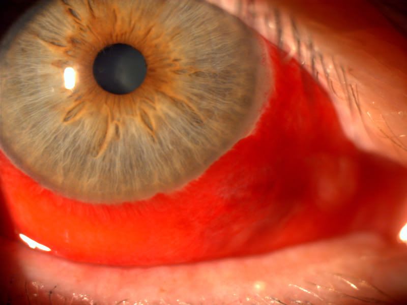
A pterygium...these are associated with years of unprotected sun exposure. So yep, wear your sunglasses.
BIG spot in the middle=Corneal ulcer due to Contact lens abuse.
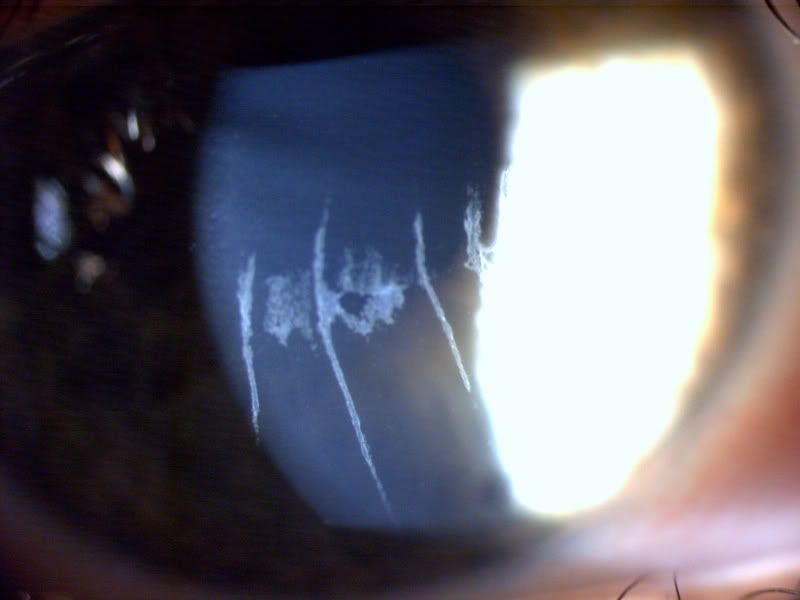
Same eye after 2 months of antibiotic treatment and a scar....
A fine diffuse irritation of the cornea, called keratitis.
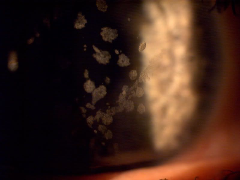
OK, cool story. Young patient comes in with this eye. Its a rare corneal dystrophy that I have never seen before and only studied about. He tells me his mother has it. Blotchy spots on the cornea, actually sees quite well.
4 months later, another patient walks in looking like this. My jaw drops and I tell the patient that I only seen it once before. Patient says, "Yeah, you prob saw my cousin" and drops his name. They live in totally different towns, different names. (I had no idea and would have thought it was simply chance that I saw 2 cases of this. Some docs go their whole life and never see one!)
Another case of contact lens abuse. My first case treating a MRSA infection in the eye. So I'll say it again, don't sleep in your contacts, ESPECIALLY if you work in a healthcare setting! THis case doesnt look as bad as the previous one above but took 3 months to treat.
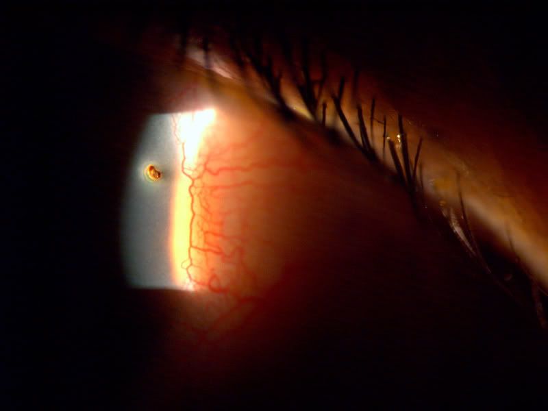
A tree branch to the eye.. the green line is due to flourescein added to dye, mark and measure it. The brown behind it its the shadow of the laceration against the iris.
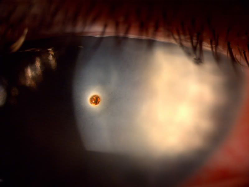
Taken this week..... He comes in, all is well, life is good, I look at his eye and Whoa, dude, you have RUST in your eye.....dont you feel it??!? He said no!
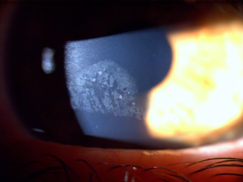
THis gent came in after "something popped in my eye" as told by an interpreter. The man repaired musical instruments. All that white haze is not artifact. It was cloudy white and he could not see anything but foggy lights. 2 weeks later, after removal, he was 20/20 with crystal clear corneas..
Ah, my favorite.....I call his one Freddy Krueger. 5 year old girl comes in with mom on a Thursday morning emergency. She has a big smile on her face, very sweet, no tears. I ask "What's wrong mom. Is there an emergency?" "I took her out of school b/c she reached up to grab a box on a shelf at school and safety scissors fell into her eye." Now, remember this girl is smiling, happy, her eye wasnt even red....and yet
This girl was once and for all proof that woman rule! Amazing, freakish pain threshold she had! 23 hours later, she was 100% resolved.
-darwin- corneal burn, secondary to a woman curling her lashes with an iron...
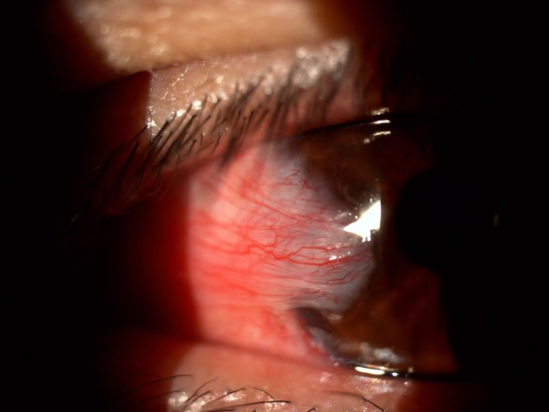
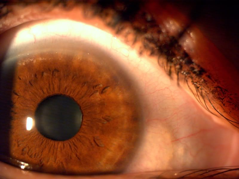
top part of the cornea, that slight haze....a potential sight of elevated cholesterol. Also, to the right of the iris, on the white part...see how there is a slight off white yellowish bump? That is a pinguecula, due to excess UV exposure.
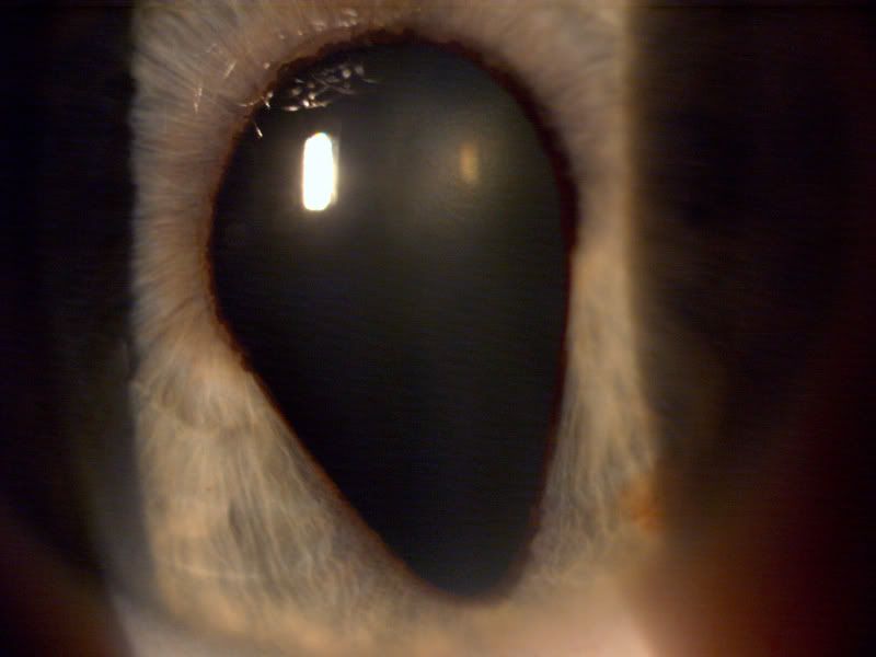
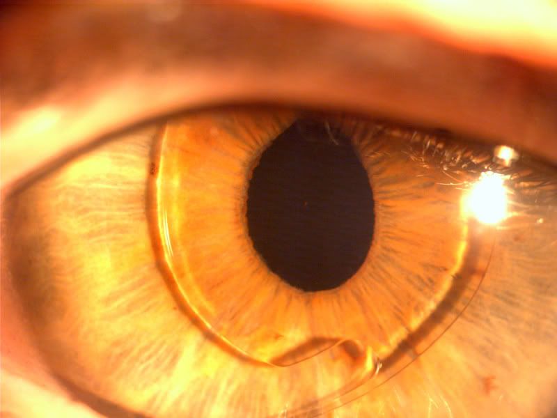
Moving back into the eye, now focused on the iris. A developmental anomaly called coloboma. The Iris never fully formed.
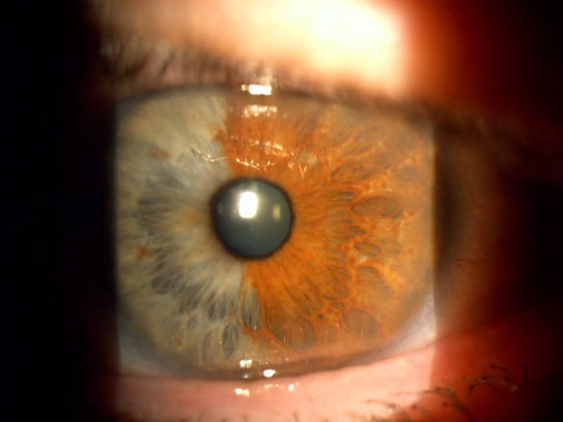
iris polychromia....genetic trait, having more than one color in an iris.
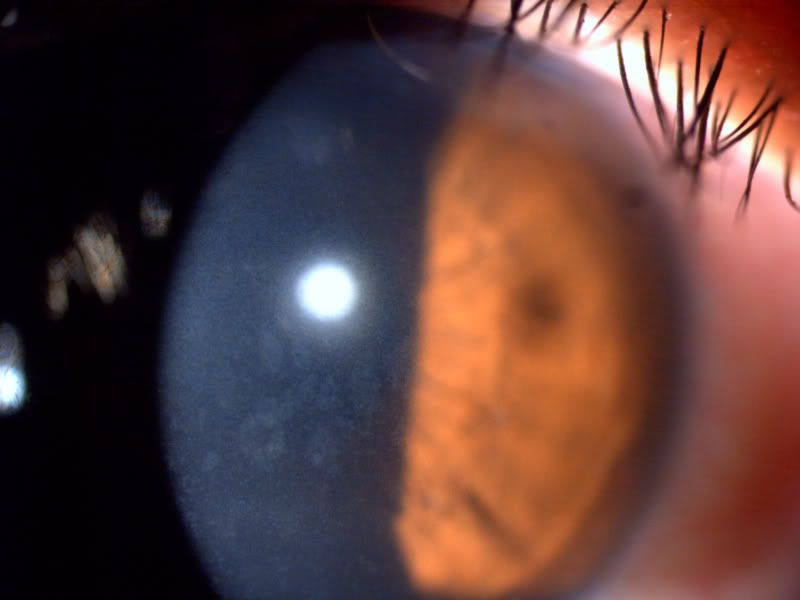
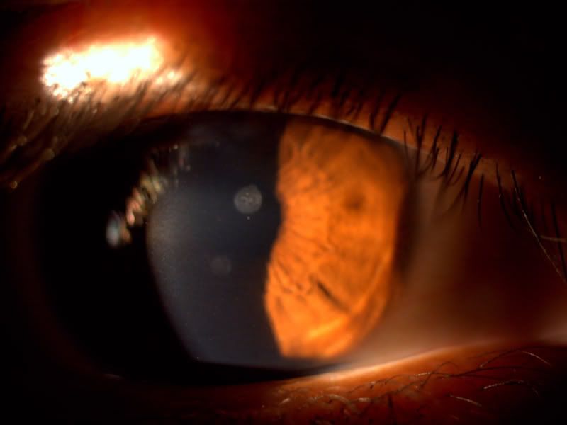
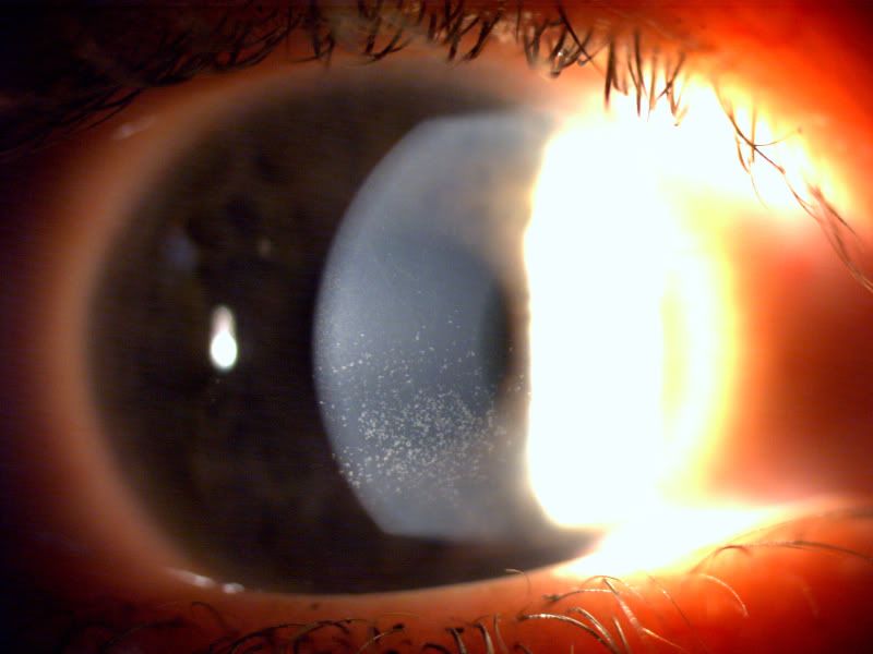
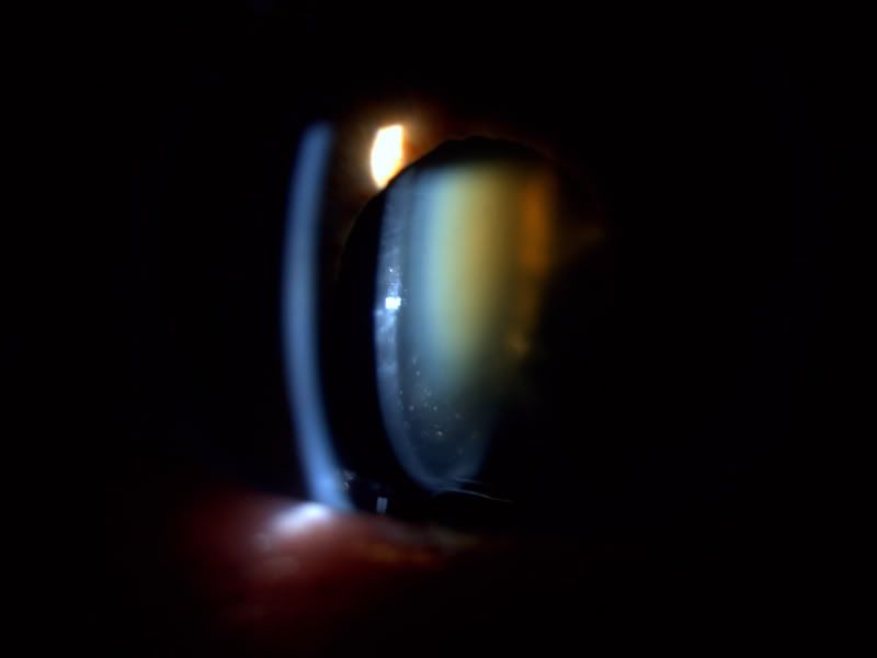
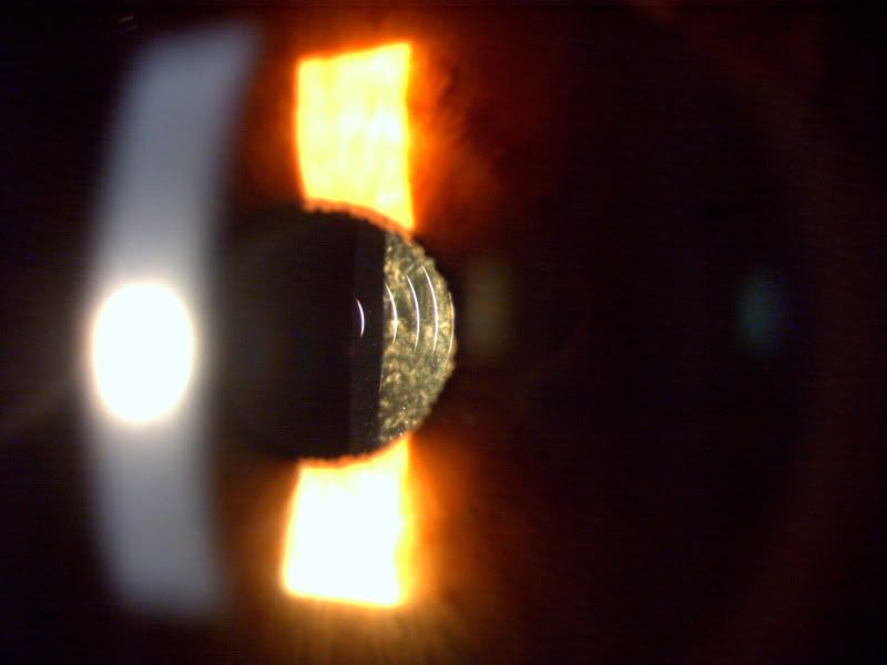
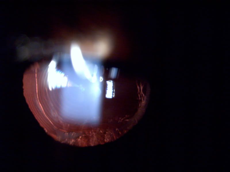
moving further back, here is a rare shot of the full breadth of a cataract. The main lens in the eye is called the crystalline lens. From the left, the first white arc is the light coming from the side and hitting the cornea. Then you have the black void, followed by the actual lens. See how it changes from a greyish white to green to brown? It should be clear/greyish. The colors are the clouding of the lens we call a cataract. Its rare to be able to see the bottom curve like that.
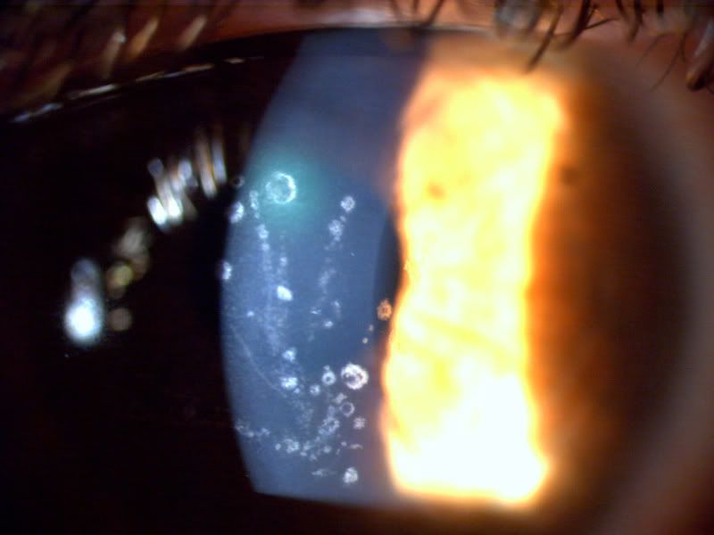
An oddball cataract...we call them tinsel or christmas cataracts b/c they are sparkly.
OK, we are still in the area of the eye where the lens sits. The following are patients who have their cataracts removed and have implants in place...
Here, a cataract was removed and replacing it is an anterior chamber intraocular lens which sits right in front of the iris. These are rare (unless you're a hack of a surgeon....) I've only seen a few of them, from patients done in the late 80s and early 90s, before my time.
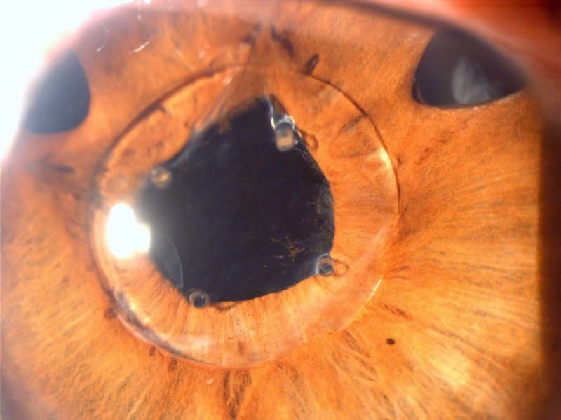
Wild one here! This man had surgery in the 80s in the Soviet Union. A unique look at their technique. Its an anterior chamber lens held by the iris. The patient describes some unique aberrations while driving....
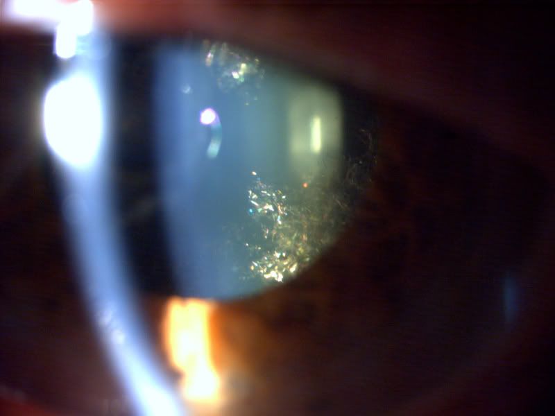
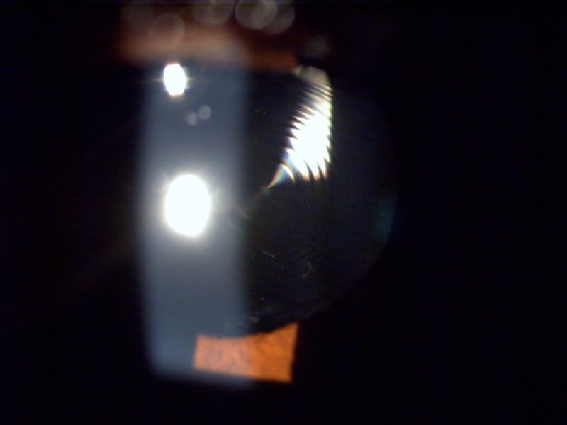
And here is some sexy new stuff. Most of you are familiar with diffractive optics lenses for cameras. We are now using them in some cataract patients!! So the cloudy lens has been replaced with a diffractive optics lens. I took a shot of a few of them, focusing on the diffractive rings. Patients like it but one nuisance is night time aberrations.
This is the same eye as above but using an imaging technique called retroillumination, where I am bouncing light off the retina to silhouette the rings. If you look closely, you can see that the ring separation is greater toward the center and the rings get incrementally closer toward to periphery.
Well thanks for looking.
I hope you found it as interesting as I did.
Note: All of these eyes, despite their appearance, are good stories. They all have happy endings. All the outcomes were 20/25 or better! So all these eyes can legally drive!
Many thanks to Fred for inspiring me to share! It's a crummy camera but it does help us get some interesting shots!


 Reply With Quote


Bookmarks